


 68 / 84

April 2016
66
ON FARM LEVEL
Integrated pest control
Several DNA-based molecular techniques are available to identify
the unknown fungal cultures. If the target fungus is unknown, a
PCR can be performed to sequence a general fungal gene, and
this sequence can be compared to thousands of fungal sequences
on an online database for identification.
When
Fusarium
root or crown rot is suspected, a species specific
PCR reaction can be performed to identify the specific
Fusarium
species. A positive identification of
Fusarium pseudograminearum
,
a common pathogen of crown rot on wheat and barley in the West-
ern Cape, could therefore take up to 14 days.
The greatest number of wheat and barley samples received by
the Disease Clinic, presented symptoms of foliar diseases. To iso-
late the causal pathogen, symptomatic leaves are surface-sterilised
and lesions excised, which is plated onto a growth medium (as de-
scribed for root fungi).
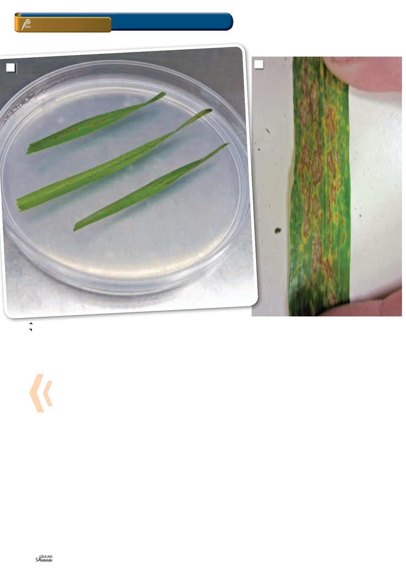
Non-sterilised leaves are also placed in ‘moisture chambers’ (petri
dishes with nutrient poor agar) (
Photo 4
), to allow the fungus to
sporulate. Fungal spores can be examined under the microscope
to identify the genus they belong to. Foliar diseases, caused by
different fungal species, may present similar symptoms. Similarly,
not all lesions and marks observed on leaves are caused by plant
pathogens (
Photo 5
).
Septoria glume blotch, caused by the fungus
Stagonospora nodo-
rum
, produce leaf lesions beginning as very dark brown flecks or
spots, sometimes with a yellow halo. These small irregular lesions
expand into oval light brown lesions with dark brown centres. These
symptoms can be confused with the high production of melanoid
pigments in some genotypes.
Pyrenophora teres
is known to cause spot and net blotch on bar-
ley. Spot blotch consists of dark-brown, circular to oval lesions,
surrounded by a chlorotic or necrotic halo. The net form lesions
have characteristic narrow, dark-brown, netlike patterns, surround-
ed by yellow or necrotic tissue. These symptoms, however, are
very similar to Ramularia leaf spot symptoms caused by
Ramularia
collo-cygni
.
The disease was first recorded in 1893, but it is only in the last
15 years that it has become recognised as an economically impor-
tant disease in Europe, Argentina and New Zealand. Ramularia leaf
spot is believed to be more widespread as it may be under-report-
ed, largely because symptoms are easily mistaken for other more
common diseases, or misidentified as physiological leaf spots.
Furthermore, this fungus is difficult to isolate from plant material
and is a slow grower on artificial media, therefore it is easily over-
looked during diagnostic procedures. Ramularia leaf spot was not
4: Symptomatic leaves are placed in a ‘moisture chamber’ to allow fungi to sporulate, thereby assisting in identification.
5: Necrotic brown, elongated and small lesions on wheat leaves from which no plant pathogenic fungi or bacteria could be isolated.
The identification of diseases on wheat and barley
5
4